
Understanding MRI Basics: Introduction to Magnetic Resonance Imaging

Magnetic resonance imaging (MRI) scans are a widely used imaging tool. MRI technology was first developed in the early 1970s. Early MRI technology proved successful in determining between normal and cancerous tissues and has continued to advance its capabilities to the modern day. In the US, it is estimated that nearly 40 million MRI scans are conducted each year.
MRI scans have proven useful in providing some of the highest-resolution images available, provide no radiation exposure (unlike x-rays or CT scans), and can image nearly any part of the body.
This article will provide details on what MRI scans are, how they work, and what to expect as a patient during an MRI scan.
What is an MRI Scan?
An MRI uses strong magnets and radio waves to create detailed images of your organs, soft tissues, bones, and even blood flow. Unlike X-rays or CT scans, MRIs don’t use radiation, making them a safe choice for most patients.
The MRI machine is a large tube that has a powerful magnet within it. The technology is complex but works in the following way:
- Our body has numerous elements within it, including hydrogen. The hydrogen atoms within our body will align themselves with the magnet in the MRI machine.
- The MRI machine produces radio waves that briefly disrupt how these hydrogen atoms are aligned.
- As the atoms realign, they emit radio signals that sensors within the MRI machine are able to detect. To the patient, this process is unfelt. They do not experience any sensation from the hydrogen atoms nor the radio waves.
- Computer technology analyzes these signals and can create images. These images are cross-sectional, meaning that the MRI machine can produce multiple images that allow a body part or organ to be seen from a variety of angles.
MRI scans are the ideal tool for viewing soft tissue, such as muscles, ligaments, tendons and organs.
The Patient Experience: What Happens During an MRI Scan?
There are several steps involved for preparing for the scan, engaging in the scan, and post-recovery.
Preparation: To prepare for an MRI scan of the abdomen or gastrointestinal system, you may be asked to avoid eating for a certain period beforehand. Once you arrive at the hospital, you'll likely change into a hospital gown and remove any jewelry, hairpins, eyeglasses or other metal items that that could interfere with the scan.
Depending upon the body part being scanned, you may be injected with a contrast. This could be considered a “dye” that will help select body tissue, organs and blood vessels show up better during the scan. In the case of MRI scans, the contrast usually contains minute particles of gadolinium. This is a rare metal that has special magnetic properties; it can briefly change the magnetism of nearby water molecules in the body, allowing the tissue or organs being scanned to have much more detailed images than if done without contrast. The contrast is safe and non-allergic for most people and flushes out of the body during urination.
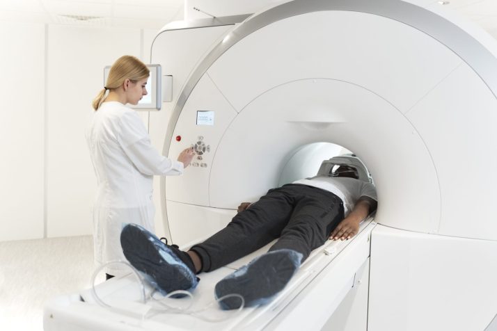
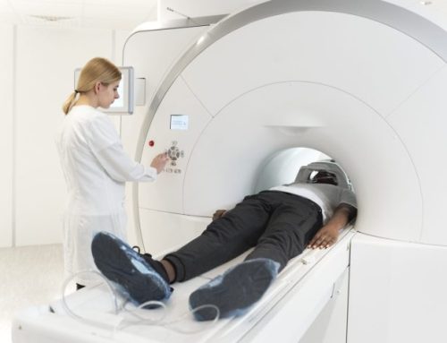
The Scan: You will lay down on a padded table that slides into the MRI scanner. You will usually lie on your back. The radiologist will provide guidance if you need to change your body position.
It is important to remain still and relaxed during the scan. If your body moves during the procedure, the process may need to be re-done, making the scanning procedure longer.
While the scanner is working, you may hear knocking or banging noises. The radiologic technician is in a neighboring control room and will be in communication with you through an intercom. There is also a camera in the MRI machine so that the technician can view you.
During the scan, the radiologist may give you instructions to either move a body part or hold your breath for a brief period to aid the imaging process. The scanning process itself causes no sensation to the patient. Depending upon the number of images needed, the process can take from 15-90 minutes.
Post-Procedure: There is no recovery period. Most patients can resume their regular activities without delay. If you received contrast, you may be encouraged to drink more fluids to help flush the contrast out of your body. This may increase your need to urinate more frequently during the rest of the day.
Common Uses of an MRI Scan
MRI scans are a powerful diagnostic technique for the following medical issues including:
- Musculoskeletal: Muscles, tendons, ligaments and cartilage can be seen in detail with an MRI scan, being able to diagnose sprains, joint injuries and spinal problems.
- Cardiovascular: Heart structure and function can be analyzed (especially damage from a heart attack) as well as the evaluation of blood vessels.
- Neurological: The brain and spinal cord can reveal conditions like strokes, tumors, multiple sclerosis, and Alzheimer’s.
- Cancer: Various cancers and their stages can be determined by an MRI scan.
- Gastrointestinal / Organs: MRIs can determine medical issues affecting the stomach, liver, kidneys, and other organs.
MRI scans play a role in monitoring medical treatment such as the effects of chemotherapy, immunotherapy or radiation therapy on cancerous tumors.
Medical planning and observation are an additional service that MRI scans provide. Surgeons are able to see detailed imagery of a surgical issue in advance, and can use that knowledge to plan the surgery, as well as anticipate potential concerns or obstacles.
RadiologyAssist: Your Partner in Radiologic Screening
We are the national leader in providing radiologic screenings to uninsured and self-pay patients. We partner with over 1000+ high-quality accredited centers nationwide, ensuring quality and convenient care with up-front transparent pricing. Our scheduling capabilities allow our patients to get their low cost MRI scans quickly, avoiding delays in diagnosis and care.
Final Thoughts
For nearly 60 years, MRI scans have been a leading diagnostic tool, safely and effectively identifying countless medical conditions. At RadiologyAssist, we make these essential scans affordable and within reach for everyone.
Blog & Healthcare News


